Furosemide is the most commonly used diuretic in critical care and is frequently used in the management of acute kidney injury (AKI). However, the benefits of furosemide administration in AKI has long been questioned and there are concerns over the possible harmful effects of furosemide including diuretic-induced AKI. To further evaluate the role of furosemide in management of AKI, let us consider a case.
Continue reading “Furosemide use in management of acute kidney injury: magic bullet or fatal blow?”Blog
A Practical Approach to Mechanical Ventilation in Small Animals
Hyperosmolar therapy in spontaneous intracerebral hemorrhage
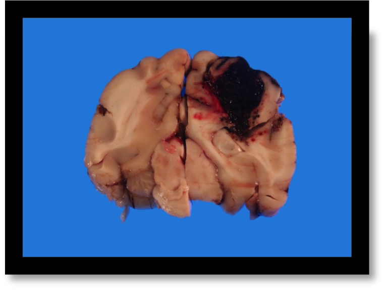
A 7 year-old female spayed Pembroke Welsh Corgi was presented to a veterinary teaching hospital for evaluation of acute onset generalized clonic-tonic seizures, obtunded mentation and petechiation. Her primary veterinarian previously saw the dog two days ago when she developed anorexia. Multiple petechiations were noted on her skin at that time. Her initial blood work revealed severe thrombocytopenia at 5-10 K/uL (RI, 190-450 K/uL). The rest of the work-up (thoracic/abdominal radiographs and tick-borne disease testing) was unremarkable, and she was prescribed prednisone for suspected primary immune-mediated thrombocytopenia (ITP). The following day, she developed two clonic-tonic generalized seizures that lasted 1-2 minutes each. Her mentation became progressively worse, and she had several seizures on the day of presentation to the teaching hospital.
Continue reading “Hyperosmolar therapy in spontaneous intracerebral hemorrhage”Myth or Fact: “Idiopathic ARDS”
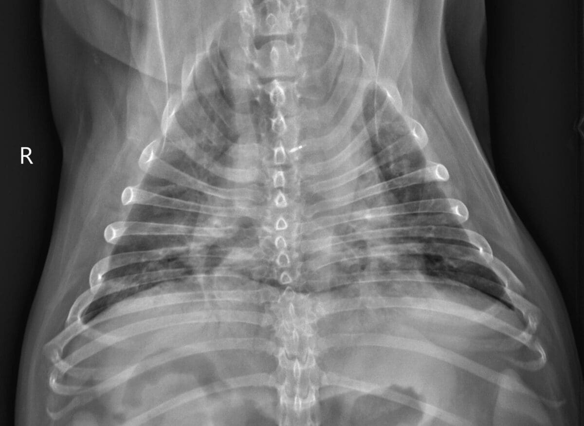
During the course of my emergency and critical care career I have seen a number of dogs that presented to the emergency room in acute respiratory distress and fulfilled almost all ARDS or VetARDS criteria (see below), however all of these patients were missing one important criterion that did not let me make a diagnosis of ARDS with confidence. This criterion is the presence of an underlying disease or risk factor predisposing them to the classic ARDS. In this article, I will discuss a so-called “idiopathic ARDS”, also known as an acute interstitial pneumonia (AIP). I will speculate that this pathologic condition remains underdiagnosed in veterinary medicine and its true prevalence in dogs and cats is unknown.
Continue reading “Myth or Fact: “Idiopathic ARDS””Venous pCO2 in Shock
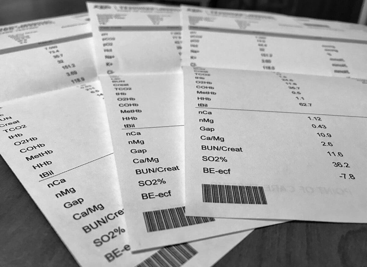
A 3 year-old neutered male domestic shorthaired cat was rolled out of an OR after the chylothorax surgery (cysterna chyli ablation, pericardiectomy, and thoracic duct ligation) as well as the surgical correction of congenital peritoneopericardial diaphragmatic hernia (PPDH). An extensive pleural fibrosis was noted during surgery due to the suspected chronicity of the chylous effusion. A unilateral small-bore chest tube was placed into one of the hemithoraces at the conclusion of the surgical procedure. The intraoperative anesthesia monitoring was complicated by the inability to obtain indirect blood pressure measurements during the second half of the procedure despite the presence of otherwise stable monitoring parameters including end-tidal CO2. No significant blood loss was noted during the surgery.
Continue reading “Venous pCO2 in Shock”


