As emergency veterinarians, one of the most critical decisions we face is whether to recommend humane euthanasia in canine patients with spontaneous (non-coagulopathic) hemoperitoneum based solely on CT results. A recent study shed light on the limitations of CT imaging in distinguishing between benign and malignant lesions in such cases (Parry et al. JVECC 2023). Understanding the study’s findings is crucial for making well-informed decisions and providing optimal care for our canine patients. In this blog post, we will explore the study’s results, particularly the concerning frequency of benign lesions being misinterpreted as malignant, and discuss the implications for our decision-making process.
Unreliable distinction between Benign and Malignant Lesions
The study’s results suggest that CT should not be solely relied upon to distinguish between benign and malignant lesions in cases of hemoperitoneum before surgical stabilization and histopathology. One striking finding is that radiologists frequently misinterpreted benign lesions as likely malignant. In fact, one out of six benign lesions (16%) and four out of six benign lesions (67%) were mistakenly classified as malignant by radiologists 1 and 2, respectively. This variability in image interpretation among radiologists highlights the inherent challenges in using CT as a definitive diagnostic tool for these cases.
Imaging Characteristics Overlap
Previous descriptions of intraabdominal masses on CT have provided limited insights, with common imaging markers failing to show a significant association with the histological diagnosis in the study. Both malignant and benign lesions demonstrated significant overlap in their imaging characteristics, further complicating the differentiation process.
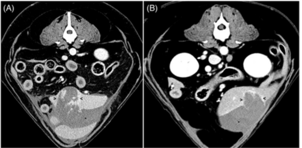
Figure 1. CT comparison between splenic hematoma (A) and splenic hemangiosarcoma (B). Arrowheads indicate the lesions. Hemorrhagic peritoneal fluid is denoted by asterisks.
Markers of Malignancy
Markers traditionally associated with malignancy, such as lesion diameter and intralesional contrast enhancement patterns, were not consistently indicative of malignancy in this study. Lesion diameter, although cited as a possible marker for malignancy in various studies, did not show a significant difference between benign and malignant lesions. Similarly, contrast enhancement patterns did not reliably distinguish between the two categories, potentially due to the predominance of single-phase contrast studies in the current study.

Considering Multifactorial Decision-Making
Given the limitations and challenges associated with CT imaging in these cases, emergency veterinarians should adopt a multifactorial approach when determining the best course of action. Relying solely on CT results to make euthanasia decisions may lead to misdiagnosis and unnecessary loss of life. Instead, emphasize the importance of surgical stabilization and histopathology in achieving a definitive diagnosis.
Study Limitations
- Low Sample Size: The study had a small sample size, which increased the risk of type 2 error. This might have affected the ability to detect statistical differences between the two groups concerning CT findings. A larger sample size, possibly through a multicenter study, would have been beneficial.
- Unblinded Radiologists: Both radiologists were aware of the dogs’ signalment when interpreting the abdominal CT studies, which could have introduced bias into their interpretations. However, since the mean age in both groups was not significantly different, the impact of this bias is deemed negligible.
- Lack of Thoracic Imaging: Reviewers did not have access to thoracic imaging when interpreting abdominal CT studies. This design choice aimed to minimize bias towards a malignant diagnosis after identifying pulmonary nodules. However, in real-life situations, radiologists have access to all available imaging results, which could affect the interpretations. Nonetheless, the study authors believe this design did not significantly impact the final results, as only a few dogs showed detectable lung metastases on thoracic imaging.
The Bottom Line
As emergency veterinarians, we face complex decisions daily, and euthanizing canine patients with spontaneous hemoperitoneum is undoubtedly one of the most challenging. The recent study’s findings remind us of the limitations of CT imaging in distinguishing between benign and malignant lesions in these cases. Rather than relying solely on CT results, we should prioritize surgical stabilization and histopathology to achieve a definitive diagnosis.

References:
Parry, M. E., Vallone, J. M., Gremillion, C. L., Wustefeld-Janssens, B. G., & Yankin, I. (2023). Retrospective evaluation of the diagnostic utility of computed tomography in dogs with nontraumatic hemoperitoneum: 26 cases (2015-2020). Journal of veterinary emergency and critical care (San Antonio, Tex. : 2001), 10.1111/vec.13310. Advance online publication. https://doi.org/10.1111/vec.13310