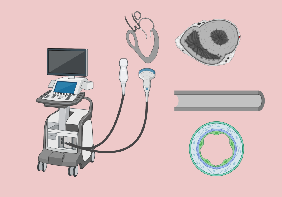
Lung and Pleural Space Ultrasound

Lung and pleural space point-of-care ultrasound is usually performed in sternal recumbency unless a dog cannot tolerate this position. In this scenario, the patient can be positioned in right or left lateral recumbency to evaluate all lung fields. The Pleural and Lung Protocol (PLUS) is my preferred method to evaluate the pleural space and lung parenchyma (Boysen et al. 2022).

Continue reading “Lung and Pleural Space Ultrasound”

Should you recommend euthanizing canine patients with spontaneous hemoperitoneum based on CT results alone?

As emergency veterinarians, one of the most critical decisions we face is whether to recommend humane euthanasia in canine patients with spontaneous (non-coagulopathic) hemoperitoneum based solely on CT results. A recent study shed light on the limitations of CT imaging in distinguishing between benign and malignant lesions in such cases (Parry et al. JVECC 2023). Understanding the study’s findings is crucial for making well-informed decisions and providing optimal care for our canine patients. In this blog post, we will explore the study’s results, particularly the concerning frequency of benign lesions being misinterpreted as malignant, and discuss the implications for our decision-making process.

Continue reading “Should you recommend euthanizing canine patients with spontaneous hemoperitoneum based on CT results alone?”

Assessment of the intravascular volume status with point-of-care ultrasound

Assessment of the intravascular volume status is a critical skill for emergency veterinarians. Point-of-care ultrasonography (POCUS) is emerging as a valuable, noninvasive, cage-side diagnostic tool for objective evaluation of physiologic and hemodynamic parameters related to volume status, fluid tolerance, and responsiveness. Rapid cage-side sonographic evaluation can obtain qualitative data on cardiac function even when performed by non-cardiologists. POCUS involves answering focused clinical questions using cage-side ultrasonography and increases the sensitivity of the conventional physical examination.

That said, POCUS findings should be interpreted in conjunction with other clinical parameters—such as conventional perfusion parameters (heart rate, mentation, body temperature, mucous membrane color, CRT, pulse quality), urine output, blood pressure, and body weight —and not viewed as an alternative to physical examination or standard imaging studies.

In this blog post, I will describe the most commonly used views to assess volume status in dogs and cats: focused sonographic assessment of the heart (cardiac POCUS) and caudal vena cava (CVC POCUS).

Continue reading “Assessment of the intravascular volume status with point-of-care ultrasound”

Pulmonary thromboembolism in dogs and cats: to lyse or not to lyse? 

An 8-year-old neutered male dachshund was presented to the emergency department for evaluation of respiratory distress. The dog was previously diagnosed with a heartworm disease and was currently undergoing treatment with a “slow kill” method. Physical examination revealed tachycardia at 160/min, right-sided pulmonary crackles, and tachypnea with increased respiratory effort. The point-of-care blood work showed hyperlactatemia at 4.5 mmol/L, PCV/TS of 53%/7.8 g/dl, mild metabolic acidosis, normal electrolytes and renal values. The arterial blood pressure (Doppler) was at 130 mm Hg. 

As the dog was getting flow-by oxygen, an abbreviated thoracic point-of-care ultrasound (T-POCUS) was performed (see below). 

Continue reading “Pulmonary thromboembolism in dogs and cats: to lyse or not to lyse? “

Radiographic confirmation of NG/NE tube placement in dogs and cats: Is it truly foolproof?

Nasogastric and nasoesophageal tubes are commonly used in small animal practice for short-term enteral nutrition. They are easy to place without general anesthesia, inexpensive and can be used for gastric decompression and drug administration.

Unfortunately, feeding tube misplacement happens much more frequently than the number of cases described in the literature. Although rare, misplacement may occur in advanced tertiary veterinary hospitals despite a strict adherence to the institutional policies and radiographic confirmation by board-certified veterinary radiologists (Rodriguez-Diaz JAAHA 2021).

Continue readingRadiographic confirmation of NG/NE tube placement in dogs and cats: Is it truly foolproof?