Nasogastric and nasoesophageal tubes are commonly used in small animal practice for short-term enteral nutrition. They are easy to place without general anesthesia, inexpensive and can be used for gastric decompression and drug administration.
Unfortunately, feeding tube misplacement happens much more frequently than the number of cases described in the literature. Although rare, misplacement may occur in advanced tertiary veterinary hospitals despite a strict adherence to the institutional policies and radiographic confirmation by board-certified veterinary radiologists (Rodriguez-Diaz JAAHA 2021).
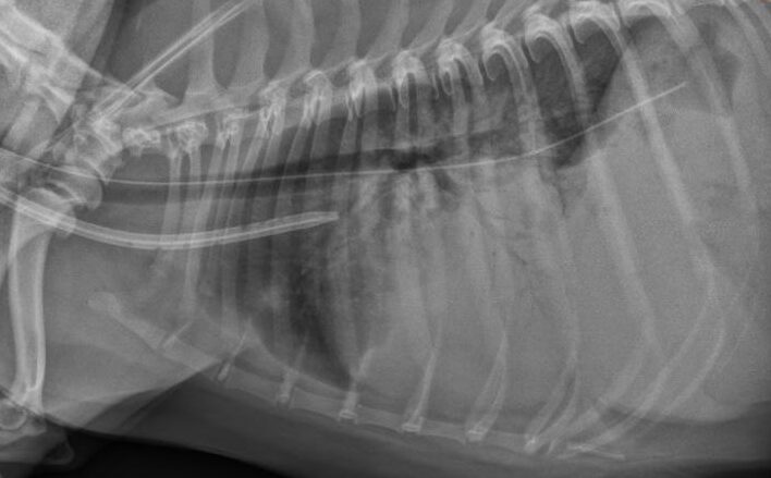
Thoracic radiography is generally considered the “gold standard” method of confirmation, however various institutions use different radiographic views for the tube placement confirmation, which, in my opinion, are not equal. Unfortunately, the most commonly used approach is to take a single standard right or left lateral thoracic view. I personally witnessed that this approach may lead to erroneous interpretation by even experienced imagers as being correctly placed in the gastrointestinal tract.
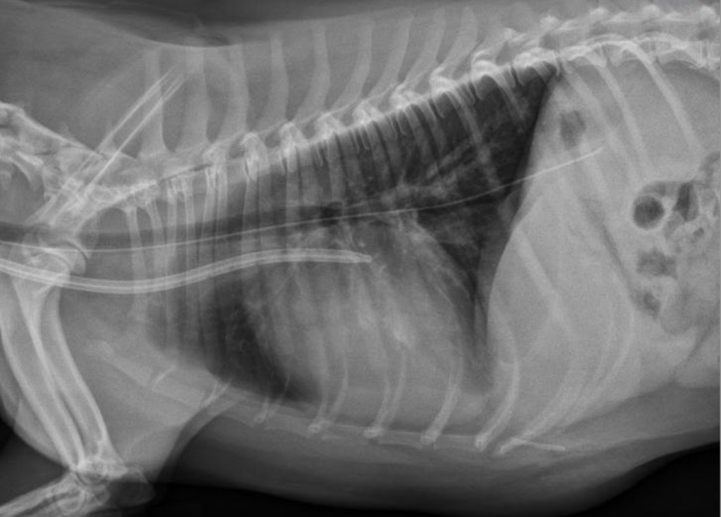
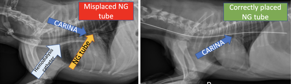
I feel that superimposition of the tube with stomach may be a primary contributor to misinterpretation because there is a significant overlap of caudal lung fields with the gastric lumen. Therefore, the location of the end of the feeding tube should NOT be used as the main criterion for determining correct placement.
What is the alternative to the single lateral thoracic view?
The most reliable results could be achieved by adhering to one of the three radiographic protocols below:
- Two-view thoracic radiographs (lateral + VD or DV) with the lateral view incorporating lateral neck and nasopharynx. This is the ideal method of radiographic confirmation of nasoenteric tubes (see explanation below).
- One lateral thoracic view incorporating lateral neck and nasopharynx. This method will dramatically increase the accuracy of radiographic confirmation; however, it lacks an additional VD view that will make this approach even more reliable.
- One lateral thoracic view with positive or negative (air) contrast study performed after obtaining plain radiographs suggestive of correct placement. This is my least favorite approach because it requires additional administration of contrast media, which is not 100% safe when the tube is misplaced.
So, what makes the protocol #1 ideal?
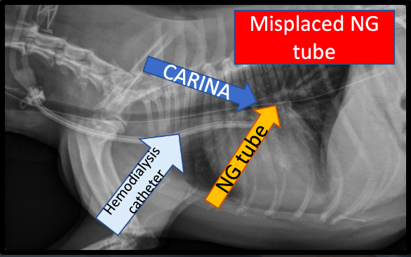
First of all, correctly placed NG/NE tubes are always located dorsal to the trachea in the region of the pharynx and, therefore, I highly recommend including this region when trying to confirm the tube placement.
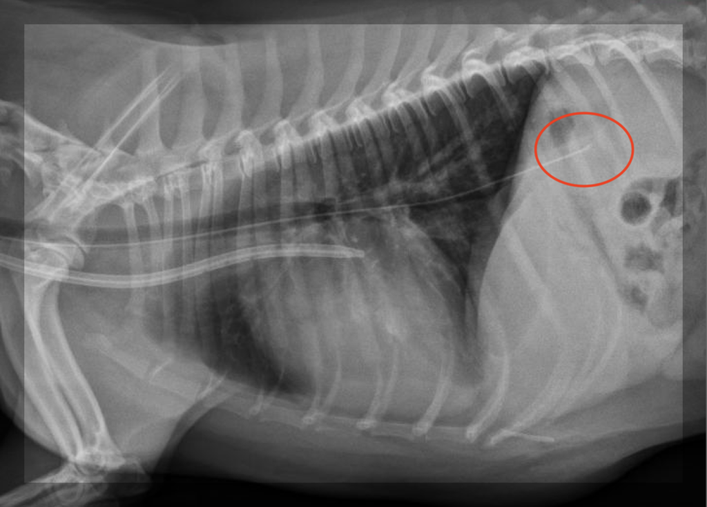
An orthogonal view (VD or DV) may provide you with extra confidence that the tube was placed correctly because in this case the tube will remain at midline in the region of caudal mediastinum whereas it will follow a caudal mainstem bronchus to the periphery (off of the midline) with incorrect placement. However, an incorrectly placed tube may still remain near midline in rare cases, without following the caudal mainstem bronchi to the periphery, as inadvertent placement into the accessory bronchus was documented in a few cases.
Finally, incorrectly placed feeding tubes tend to superimpose with the carina while correctly placed tubes will go above the carina, which appears to be a reliable indicator of a feeding tube placement as well.
The Bottom Line
Taking only one standard single lateral thoracic view is not foolproof for radiographic confirmation of nasoenteric tube placement. Two-view thoracic radiographs with the lateral view incorporating lateral neck and nasopharynx will significantly increase the accuracy of this test.
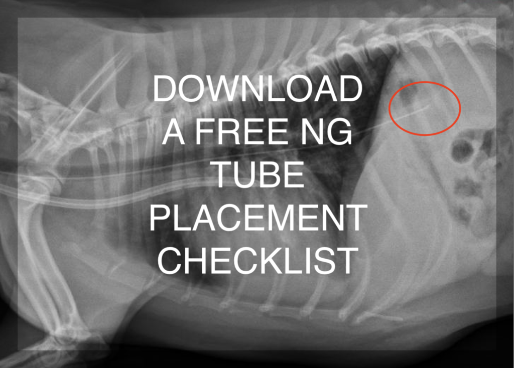
You can download the free NG/NE tube placement checklist here.
The video version of this blog post can be found here:
If you liked this video, subscribe to the VetEmCRIT Youtube Channel!
References
- Rodriguez-Diaz J, Sumner JP, Miller M. Fatal Complications of Nasogastric Tube Misplacement in Two Dogs. J Am Anim Hosp Assoc. 2021 Sep 1;57(5):242-246. doi: 10.5326/JAAHA-MS-7104. PMID: 34370835.
One thought on “Radiographic confirmation of NG/NE tube placement in dogs and cats: Is it truly foolproof?”