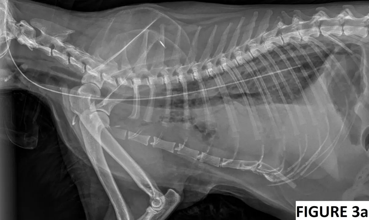
An approximately 9-year-old domestic long-haired neutered male cat was presented to his primary veterinarian for evaluation of decreased appetite, possible oral pain, and difficulty chewing food. The cat had previously undergone splenectomy and was diagnosed with a disseminated mast cell tumor that was controlled by palladia and prednisolone.
Continue reading “Is it possible to misplace an esophagostomy tube into the mediastinum?”Author: Igor Yankin
Decoding Calcium Levels in AKI Cases
A 3-year-old castrated male domestic shorthaired cat was presented to the emergency service for evaluation of acute severe lethargy, anorexia and vomiting. He has been strictly outdoors and was previously in good health. His last annual examination and blood work were performed 2 weeks ago, and they were within normal limits.
Today, his physical examination showed a body temperature of 97F (36.1C), heart rate of 140 beats per minute, respiratory rate of 44 breaths per minute, stuporous mentation, poor femoral pulses, pale pink mucous membranes, unkempt haircoat and dehydration of at least 7-8%. An ECG, venous blood gas, and thoracic/abdominal point-of-care ultrasound (POCUS) were performed during the primary survey as the intravenous catheter was being placed.
Continue reading “Decoding Calcium Levels in AKI Cases”LRS vs Citrate: Friend or Foe?
Imagine having a patient who needs simultaneous administration of a citrated blood product and Lactated Ringer’s solution (LRS), but there is only one available peripheral catheter.
Can you administer citrated donor blood or plasma and LRS at the same time?
Conventionally, this practice is discouraged because LRS is a calcium-containing fluid that may be incompatible with citrate due to the risks of calcium chelation and clot formation. Once LRS exceeds the chelating capabilities of the citrate in the stored blood that may result in clot formation, which makes perfect sense. However, as we all know, theory and practice are not the same in clinical medicine. Therefore, let’s explore the evidence to answer this very practical question.
Continue reading “LRS vs Citrate: Friend or Foe?”Dextrose:insulin ratio when treating hyperkalemia
Hyperkalemia in dogs and cats can be treated with a wide variety of methods in an emergency veterinary setting. These methods may include intravenous isotonic crystalloid fluid therapy, insulin with dextrose, sodium bicarbonate, adrenergic receptor agonists (e.g. albuterol or terbutaline) and, ultimately, treatment of the underlying condition (e.g. relief of the urethral obstruction).
Continue reading “Dextrose:insulin ratio when treating hyperkalemia”Fluid responsiveness: is it that simple?
Fluid therapy is the most commonly used initial therapeutic intervention in treatment of shock states (e.g. in hypovolemic and distributive types of shock). The approach to fluid resuscitation evolved over the last few decades. For instance, we no longer start with a full “shock dose” of fluids (60-90 ml/kg), and instead, an incremental 10-20 ml/kg boluses are preferred with frequent reassessment of the end-goal perfusion parameters. The standard therapeutic targets include improving heart rate, pulse quality, capillary refill time (CRT), non-invasive or invasive blood pressure and mucous membrane color.
But, how reliable is this approach in assessment of fluid responsiveness? Today, I will explore available evidence and diagnostic tools that can be utilized in evaluation of fluid responsiveness in veterinary and human patients.
Continue reading “Fluid responsiveness: is it that simple?”


