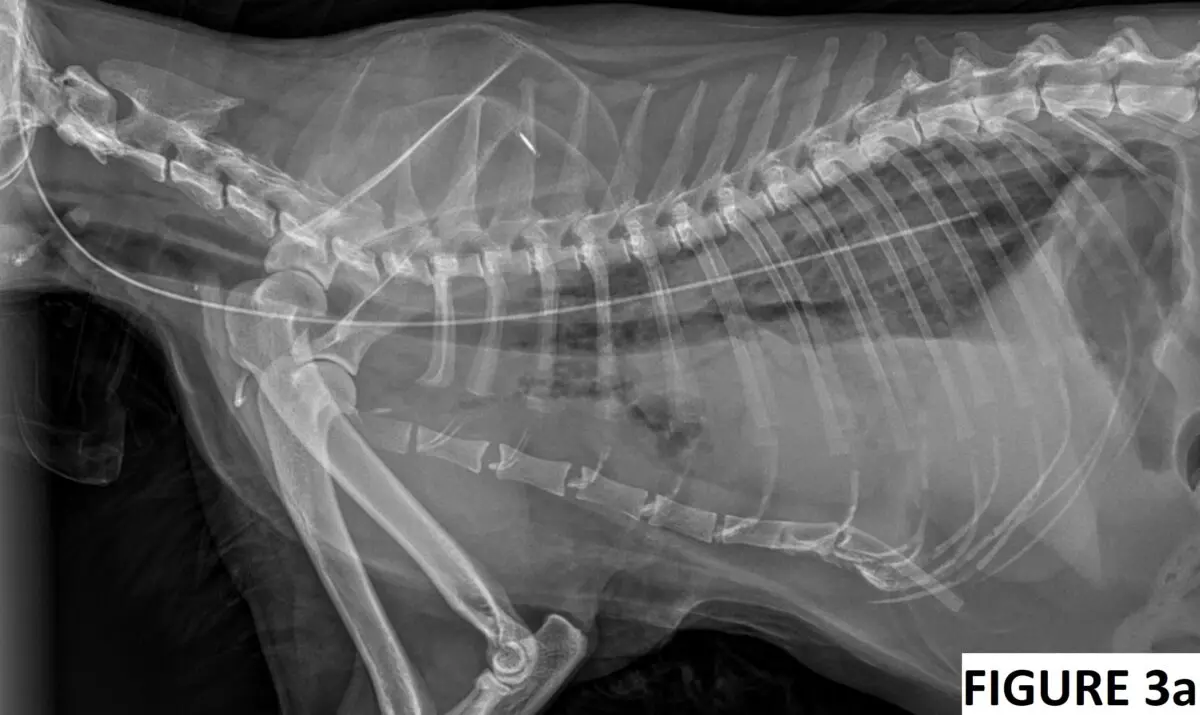
An approximately 9-year-old domestic long-haired neutered male cat was presented to his primary veterinarian for evaluation of decreased appetite, possible oral pain, and difficulty chewing food. The cat had previously undergone splenectomy and was diagnosed with a disseminated mast cell tumor that was controlled by palladia and prednisolone.
Continue reading “Is it possible to misplace an esophagostomy tube into the mediastinum?”Galleries
Decoding Calcium Levels in AKI Cases
A 3-year-old castrated male domestic shorthaired cat was presented to the emergency service for evaluation of acute severe lethargy, anorexia and vomiting. He has been strictly outdoors and was previously in good health. His last annual examination and blood work were performed 2 weeks ago, and they were within normal limits.
Today, his physical examination showed a body temperature of 97F (36.1C), heart rate of 140 beats per minute, respiratory rate of 44 breaths per minute, stuporous mentation, poor femoral pulses, pale pink mucous membranes, unkempt haircoat and dehydration of at least 7-8%. An ECG, venous blood gas, and thoracic/abdominal point-of-care ultrasound (POCUS) were performed during the primary survey as the intravenous catheter was being placed.
Continue reading “Decoding Calcium Levels in AKI Cases”Dextrose:insulin ratio when treating hyperkalemia
Hyperkalemia in dogs and cats can be treated with a wide variety of methods in an emergency veterinary setting. These methods may include intravenous isotonic crystalloid fluid therapy, insulin with dextrose, sodium bicarbonate, adrenergic receptor agonists (e.g. albuterol or terbutaline) and, ultimately, treatment of the underlying condition (e.g. relief of the urethral obstruction).
Continue reading “Dextrose:insulin ratio when treating hyperkalemia”Hypercalcemia in dogs with Addison’s disease
Hypoadrenocorticism (i.e., Addison’s disease) is an important differential for hypercalcemia. The etiology of hypercalcemia in hypoadrenocorticism in dogs is unclear. Hall et. al (JVIM 2023) wanted to find out the prevalence and factors associated with hypercalcemia in this population of dogs by performing a multicenter retrospective observational study at the 4 UK referral hospitals. They analyzed data from 110 dogs and found that about 34.5% of the dogs with Addison’s had either total and/or ionized hypercalcemia. The odds of hypercalcemia were increased (P < .05) in dogs with classic Addison’s (deficient in both mineralo- and glucocorticoids), higher serum creatinine, and higher serum albumin. The odds of ionized hypercalcemia were increased (P < .05) with reduced serum potassium concentration and younger age.
Continue reading “Hypercalcemia in dogs with Addison’s disease”Lung and Pleural Space Ultrasound

Lung and pleural space point-of-care ultrasound is usually performed in sternal recumbency unless a dog cannot tolerate this position. In this scenario, the patient can be positioned in right or left lateral recumbency to evaluate all lung fields. The Pleural and Lung Protocol (PLUS) is my preferred method to evaluate the pleural space and lung parenchyma (Boysen et al. 2022).
Continue reading “Lung and Pleural Space Ultrasound”