During the course of my emergency and critical care career I have seen a number of dogs that presented to the emergency room in acute respiratory distress and fulfilled almost all ARDS or VetARDS criteria (see below), however all of these patients were missing one important criterion that did not let me make a diagnosis of ARDS with confidence. This criterion is the presence of an underlying disease or risk factor predisposing them to the classic ARDS. In this article, I will discuss a so-called “idiopathic ARDS”, also known as an acute interstitial pneumonia (AIP). I will speculate that this pathologic condition remains underdiagnosed in veterinary medicine and its true prevalence in dogs and cats is unknown.
Continue reading “Myth or Fact: “Idiopathic ARDS””Tag: Pulmonary
Venous pCO2 in Shock
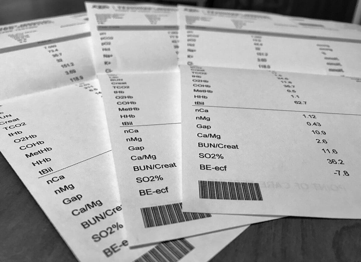
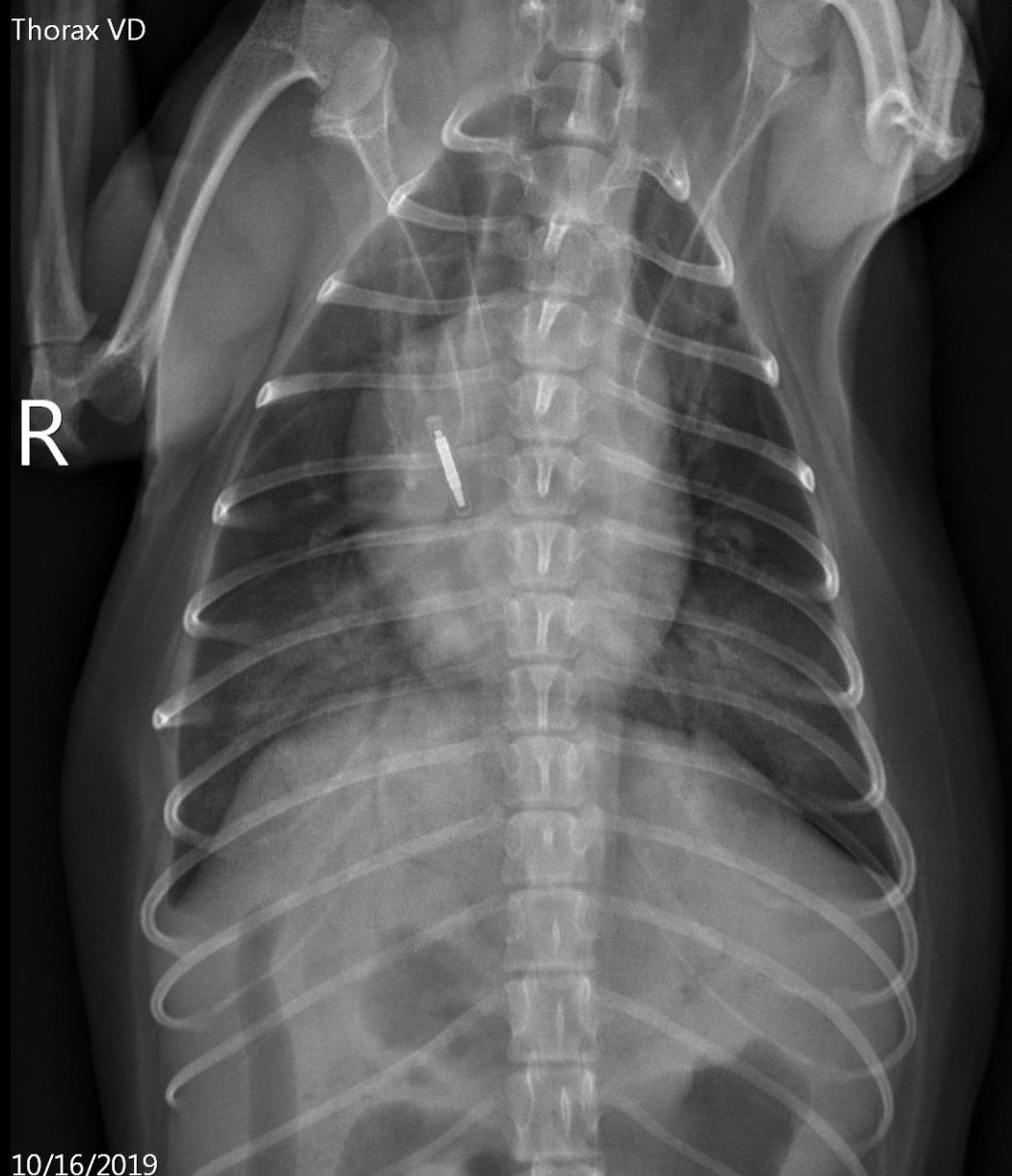
A 3 year-old neutered male domestic shorthaired cat was rolled out of an OR after the chylothorax surgery (cysterna chyli ablation, pericardiectomy, and thoracic duct ligation) as well as the surgical correction of congenital peritoneopericardial diaphragmatic hernia (PPDH). An extensive pleural fibrosis was noted during surgery due to the suspected chronicity of the chylous effusion. A unilateral small-bore chest tube was placed into one of the hemithoraces at the conclusion of the surgical procedure. The intraoperative anesthesia monitoring was complicated by the inability to obtain indirect blood pressure measurements during the second half of the procedure despite the presence of otherwise stable monitoring parameters including end-tidal CO2. No significant blood loss was noted during the surgery.
Continue reading “Venous pCO2 in Shock”TRALI or Pulmonary Hemorrhage?
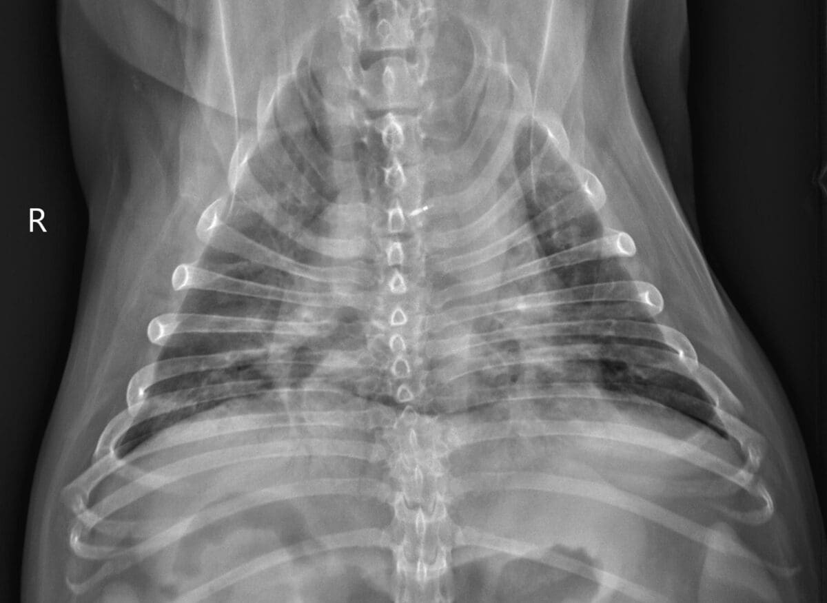
A 7 year-old spayed female miniature poodle was referred to a veterinary teaching hospital emergency service for further evaluation of severe thrombocytopenia due to a suspected immune-mediated thrombocytopenia with a platelet count of 12k/uL confirmed by a blood smear. Initial work-up included standard bloodwork, a tick-borne disease panel, thoracic and abdominal imaging. No apparent underlying causes of presumed ITP were identified. An initial physical examination showed moderate tachycardia with a HR of 150/min, strong femoral pulses, pale mucous membranes and multifocal cutaneous petechiation. The dog was eupneic and had normal bronchovesicular sounds. There was melena on rectal palpation. The patient’s PCV was 20% with TS of 5.5 g/dl.
Continue reading “TRALI or Pulmonary Hemorrhage?”