Acid-Base Analysis in a Cat

This case demonstrates the value of advanced acid-base analysis in a cat with euglycemic diabetic ketoacidosis.

Acid-base analysis in a cat can be deceptively complex, especially when traditional parameters like pH, pCO₂, and bicarbonate appear normal. In this case, advanced analysis revealed a hidden mixed disorder that would have been missed without deeper evaluation.

You are presented with a 12-year-old spayed female domestic shorthair cat with a history of injection-site sarcoma and diabetes mellitus. She is currently being treated with an SGLT2 inhibitor, a sodium–glucose co-transporter-2 blocker.

On physical examination, she was dull, dehydrated, and hypothermic.

A venous blood sample was obtained immediately after IV catheter placement. The collection was performed under strict anaerobic conditions, without struggling, and with no evidence of sampling error.

The venous blood gas panel was then analyzed, and the results are shown below.

  • pH = 7.427 (Reference: 7.244 – 7.444)
  • pCO₂ = 28.7 mmHg (Reference: 27.3 – 49.1)
  • HCO₃⁻ = 19.11 mmol/L (Reference: 15.9 – 24.7; mid-normal: )
  • Base excess (BEecf) = – 5.5 mmol/L (Reference: -10.4 to -0.5)
  • Sodium = 137.4 mmol/L (Reference: 150.5 – 157.2; mid-value: 153.9)
  • Potassium = 2.96 mmol/L (Reference: 3.11 – 4.64)
  • Chloride = 95.9 mmol/L (Reference: 113 – 123; mid-value: 118)
  • Ionized calcium = 1.15 mmol/L (Reference: 1.21 – 1.45)
  • Glucose = 135 mg/dL, or 7.5 mmol/L (Reference: 74 – 159 mg/dL = 4.1 – 8.8 mmol/L)
  • Lactate = 2.3 mmol/L (Reference: 0 – 3.3 mmol/L)
  • Creatinine = 1.25 mg/dL, or 110.5 µmol/L (Reference: 0.8 – 1.8 mg/dL = 70.7 – 159.1 µmol/L)
  • BUN = 20 mg/dL, or 7.1 mmol/L (Reference: 19 – 33 mg/dL = 6.8 – 11.8 mmol/L mmol/L)
  • Anion gap = 25.4 (Reference: 14.8 – 23.8; mid-normal: )
  • Albumin = 3.5 g/dL = 35 g/L (Reference: 2.2 – 4 mg/dL; mid-normal value: 3.1 mg/dL)
  • Phosphate =  1.3 mmol/L =  3.9 mg/dL (Reference: 3.1 – 7.5 mg/dL; mid-normal value: 5.3 mg/dL)

Now, let’s apply the advanced traditional approach to this case to perform an acid-base analysis in this cat.

Continue reading

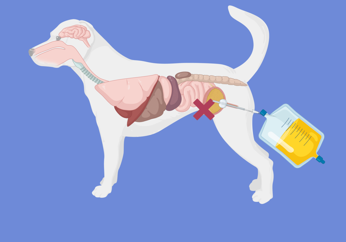
Conservative Management of Urinary Tract Rupture in Dogs and Cats: An Evidence-Based Guide for Emergency Veterinarians

Conservative, non-surgical management of urinary tract rupture is emerging as a viable alternative to surgery in select dogs and cats. This detailed review breaks down new data from several veterinary studies, offering a practical, step-by-step algorithm for emergency vets managing these challenging cases.

In this post, we’re breaking down the latest data on conservative—meaning non-surgical—management of urinary tract rupture in dogs and cats.

We’ll compare a few recent studies and walk through a practical decision-making algorithm that you can actually use in clinical practice.

Continue reading

Traumatic Brain Injury in a Cat: Hyperglycemia and suspected central diabetes insipidus

This case study of a cat with traumatic brain injury explores the pathophysiology, diagnosis, and management of post-traumatic hypernatremia, hyperglycemia, and suspected central diabetes insipidus, offering practical strategies for fluid therapy, glycemic control, and neurocritical care.

In today’s case study, we delve into the journey of Boris, a six-month-old intact male kitten who suffered a traumatic brain injury (TBI) after being kicked by a goat. His case was further complicated by hypernatremia and hyperglycemia, presenting a unique challenge for his emergency care team.

Continue reading

Point-of-care ultrasound of left ventricle in dogs and cats with shock

Learn how to visually assess left ventricular function in dogs and cats using focused cardiac ultrasound—fast, practical, and no measurements required.

In emergency situations, particularly when treating pets in shock, quickly assessing left ventricular (LV) function is crucial for determining the best course of action and accurately diagnosing the type of shock (cardiogenic vs. hypovolemic vs. distributive vs. obstructive). A fast, reliable method that emergency veterinarians can use is Point-of-Care Ultrasound (POCUS in dogs and cats with shock). This “eyeball” approach allows us to evaluate LV contractility efficiently.

In this post, I’ll walk you through the three primary components of LV function assessment using POCUS, which can be easily applied by non-cardiologists. By mastering these techniques, you’ll be able to identify whether your patient’s LV function is hyperdynamic, normal, or depressed in just a few moments.

(more…)

Blood gas analysis training for ER Vets

This blood gas analysis training for emergency veterinarians, interns, residents, and ECC or anesthesia technicians delivers a focused, interactive, and retention-driven approach to mastering acid–base and electrolyte disorders. Unlike traditional CE courses, webinars, or lectures—often long, passive, and quickly forgotten—this program uses short, targeted lessons, real-life emergency case scenarios, active learning quizzes with feedback, and a collaborative community to keep you engaged and ensure lasting knowledge.

You’ll also gain access to our Emergency Electrolyte and Acid–Base Calculator, allowing you to perform rapid, accurate supplementation calculations and full acid–base interpretation in seconds, giving you the confidence to apply what you’ve learned immediately in critical situations.

Over the last 15 years, I have attended dozens of CE events and watched hundreds of hours of webinars, only to realize a week later that I can recall only  5-10% of this information. Have you ever felt that way?

Continue reading “Blood gas analysis training for ER Vets”