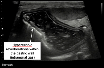
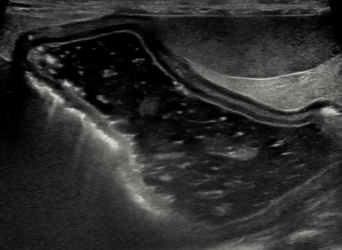
A 15 year-old neutered male Chihuahua was presented to a university teaching hospital for further evaluation of acute vomiting, anorexia, hyperbilirubinemia and elevated liver enzymes. The abdominal ultrasound was suggestive of acute severe pancreatitis resulting in extrabiliary bile duct obstruction. In addition, the gastric wall contained intramural gas consistent with gastric pneumatosis (Figure 1).
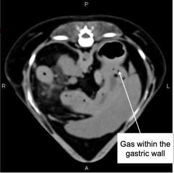
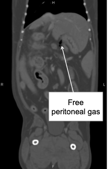
A conservative medical management was initiated that included pain management, fluid therapy and nasogastric tube feeding. Despite the outlined therapy, the dog’s severe hyperbilirubinemia (15 mg/dl, RI 0-0.5 mg/dl) persisted and he remained anorexic. The repeat complete blood cell count revealed worsening severe neutrophilic leukocytosis (WBC=71 K/uL, RI 5-15 K/uL). An abdominal CT was performed to further evaluate the hepatobiliary tract and rule out other causes of persistent physical and clinicopathological abnormalities. The CT showed moderate amount of gas within the gastric wall, small amount of free peritoneal gas, and ongoing severe pancreatitis (Figure 2 and 3).
Due to the concerns with respect to possible gastric wall necrosis leading to the gastric wall perforation in conjunction with persistent extrabiliary duct obstruction resulting in refractory hyperbilirubinemia, the decision was made to perform an exploratory laparotomy. Upon surgical exploration, no source of free peritoneal gas was identified and the serosal surface of the gastric wall appeared normal; the pancreas looked edematous and inflamed. The liver and pancreas were biopsied prior to the abdominal closure. Intraoperatively, the dog developed severe arterial hypotension refractory to volume resuscitation and requiring an initiation of norepinephrine infusion. The dog’s severe hypotension persisted after the completion of the surgery and resulted in death 24 hours after the conclusion of the surgical exploration. The histopathologic evaluation of the pancreas was consistent with severe pancreatitis.
Case Discussion
In this post, I will attempt to explore the significance of gas within the gastric wall and will propose a diagnostic and treatment approach to this population of patients.
Gastric pneumatosis is a generalized term that includes any cause of air in the wall of the stomach (Johnson et al, 2011). Human medical literature further subdivides intramural gastric wall gas into two distinct clinical entities: gastric emphysema and emphysematous gastritis.
Gastric emphysema is the dissection of air into the gastric wall through an insult caused by damage to the gastric mucosa (Zenooz et al, 2007). Emphysematous gastritis is a rare form of gastritis due to invasion of gas producing organisms (Zenooz et al, 2007). It is believed that gastric emphysema is a relatively benign and potentially self-limiting condition (Majumder et al, 2012), whereas emphysematous gastritis typically leads to systemic signs of toxicity (i.e. sepsis) and has a higher mortality rate (Seaman et al, 1967).
Etiology and pathophysiology
When gastric pneumatosis is secondary to gastric pathology, some breach in the integrity of the stomach wall is important in the pathophysiology. Massive gastric distention causing increased intraluminal pressure and gastric ischemia may lead to loss of mucosal integrity and luminal gas extension into the stomach wall (Moosvi et al, 1990).
Jones et al. (JVECC 2024) recently published the largest veterinary case series (26 dogs and 4 cats) on gastrointestinal pneumatosis. The most common sites of intramural gas accumulation were the stomach (19 cases), followed by the colon (8 cases) and small intestine (2 cases). Common diagnoses that were associated with gastrointestinal pneumatosis included gastric dilatation and volvulus (5 cases), acute hemorrhagic diarrhea syndrome (4 cases), and gastrointestinal ulceration (4 cases).
Includes the following bonuses:
Video lesson on treatment of refractory hypokalemia;
Parenteral potassium supplementation checklist [PDF];
Acid-base analysis worksheet [PDF];
Certificate of achievement (RACE);
Reported gastric etiologies of gastric pneumatosis in human and veterinary literature
1. Raised intragastric pressure
- GDV
- Gastroscopy
- Intestinal, pyloric obstruction
2. Trauma
- Injuries from endoscopic biopsy
- Nasogastric tube placement
- Blunt trauma to the gastric wall
- Gastric bezoar and foreign bodies
3. Severe vomiting
4. Gastric wall ischaemia, inflammation, infiltration
- Gastric neoplasia
- Gastric ulceration (including secondary to NSAIDs, steroids)
- Hydrogen peroxide ingestion
- Gastritis
- Bacterial gastric wall infection
- Thromboembolism
- GDV
- Idiopathic
In addition to gastric pathology, gas can travel to the stomach wall from extra-gastric sources, including the small bowel, colon, and even from the lungs dissecting along the interstitial planes. In these cases, the gastric mucosa could be intact (Johnson et al, 2011; Tuck et al, 1987).
Reported extragastric etiologies of gastric pneumatosis in human and veterinary literature
1. Dissection of air from the mediastinum
- Pneumothorax;
- Pneumomediastinum;
2. Dissection of air from intra-abdominal structures
- Perforated gangrenous cholecystitis;
- Biliary stent-related perforation;
- Pneumatosis intestinalis, coli;
- Small bowel or colon ischemia;
3. Miscellaneous/other
- Pancreatitis;
- Mesenteric artery syndrome;
- Cholangiocarcinoma.
Diagnostic dilemma
Distinction of patients whose gastric pneumatosis is due to emphysematous gastritis or ischemia, as opposed to more benign gastric processes, would be extremely valuable (Johnson et al, 2011). The differentiation of these two clinical conditions relies on the patient’s clinical presentation, clinicopathologic findings, assessment of predisposing conditions and diagnostic imaging (especially, CT). That being said, currently, there are no clear diagnostic protocols available that would guide clinicians on differentiation between the two distinct entities. Due to a significant overlap, none of the diagnostic modalities will provide a clinician with a definitive diagnosis.
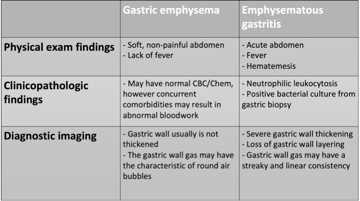
The lack of highly specific and sensitive testing makes it very hard to decide if a patient needs surgical exploration or medical management will suffice. Multiple human case series and a few veterinary case reports published over the last 10-20 years suggest that surgery can be avoided even in patients with emphysematous gastritis in the absence of bowel perforation, necrosis or ischemia. There have been reports of successful conservative treatment of this condition without any operative intervention (Paul et al, 2010). A gastrectomy is often necessary when expectant medical management fails or gastric perforation/necrosis is confirmed.
Free peritoneal gas
Evidence of pneumoperitoneum was described in the dog I presented in the beginning of this post. This gas likely resulted from diffusion from the gastric wall to the peritoneum since no evidence of viscous organ perforation or external trauma was evident in that dog.
Pneumoperitoneum has also been reported in human and veterinary patients with gastric emphysema and without gastric perforation and is not always considered to be a characteristic warranting surgery (Spektor et al, 2014; Muthukumarasamy et al, 2010; Thierry et al, 2019). In Jones et al. (JVECC 2024) paper, pneumoperitoneum was concurrently identified in 7 of 30 cases (23%), peritoneal effusion in 12 of 30 cases (40%), and hepatic portal vein gas in 4 cases (13%).
At the same time, there is always a possibility that the patient’s gastric pneumatosis and pneumoperitoneum is caused by gastric perforation and necrosis.
In a recent veterinary case report (Gordon et al. JVECC 2024), authors described a dog with hydrogen peroxide ingestion (3% solution, 10-20 ml/kg). Abdominal radiographs revealed pneumoperitoneum, gastric pneumatosis, and hepatic venous gas. The dog was managed conservatively with supportive care and oxygen therapy. Repeat radiographs 6 hours later showed complete resolution of all gas within the stomach wall, peritoneal space and hepatic veins. An abdominal ultrasound was performed during hospitalization, and it revealed severe gastric wall thickening. The upper GI endoscopy confirmed severe gastric mucosal necrosis without evidence of deeper ulceration. The dog was ultimately discharged after 5 days of hospitalization and continued to do well at home with medical management alone. Recheck ultrasound 5 weeks later showed normal gastric wall appearance.
Diagnostic and treatment approach to dogs and cats with gastric pneumatosis
Due to the rarity of gastric pneumatosis in the absence of GDV, there is a lack of human or veterinary publications that would focus on the diagnostic approach or provided the perspective that would be most helpful for safe and expeditious recognition and management of this condition. I would like to provide my personal perspective on treatment strategies for gastric pneumatosis that is based on human and veterinary literature review (Figure 4).
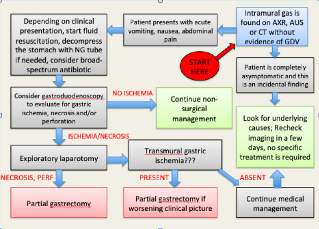
AXR – abdominal radiographs; AUS – abdominal ultrasound; GDV – gastric dilatation volvulus;
The algorithm starts when a patient is diagnosed with intramural gas that is found on abdominal imaging (radiographs, ultrasound or CT).
If a patient presents with gastrointestinal signs such as nausea, vomiting, cranial abdominal pain, abdominal distension and possible hemodynamic instability, initial interventions should include fluid resuscitation, nasogastric tube placement to decompress the stomach if it is distended, appropriate hemodynamic monitoring, pain medications, and broad-spectrum antibiotics to cover mostly gram negatives and anaerobes.
Urgent esophagogastroduodenoscopy (EGDS) should be performed to evaluate gastric mucosa for evidence of ischemia, necrosis and perforation (if that have not been identified on initial abdominal imaging). Exploratory laparotomy is indicated if there is evidence of mucosal ischemia/necrosis within the stomach. Gastric mucosa specimens should be sent for pathology, culture, and sensitivity. If exploratory laparotomy identifies transmural gastric ischemia/necrosis, partial gastrectomy should be performed. If the gastric serosa looks healthy, the ischemia/necrosis likely affected only mucosa. In this case, medical management should be continued without partial gastrectomy. Close observation is still indicated as ischemia of the gastric wall may develop in a delayed fashion.
If medical management was elected or a patient doesn’t show any signs of illness, associated conditions and underlying causes of intramural gastric gas should be investigated by obtaining a detailed history and performing further diagnostics.
Human and some veterinary case reports suggest that it might be safe to treat patients with even emphysematous gastritis for bacterial infection, in the absence of any systemic toxicity, shock, or peritonitis and provided that endoscopy can demonstrate limited involvement of the mucosa with acute inflammation and no necrosis (Matsushima et al, 2015; Thierry et al, 2019).
Gastric pneumatosis in GDV
According to a retrospective study by Fischetti et al (VRU 2004), 22/243 (9.1%) dogs with GDV had radiographic evidence of pneumatosis on at least one view and the gas within the wall was consistently linear to curvilinear, parallel to the wall, and present within the body or fundus of the stomach. Gastric pneumatosis was insensitive (14.1%) for ruling out gastric necrosis yet relatively specific (92.7%) for ruling in gastric necrosis in dogs with GDV. When pneumatosis was identified on a radiograph of a dog with GDV, there was a 40.9% chance (positive predictive value based on incidence of gastric necrosis 26.6%) that the dog will require gastric resection at surgery. Conversely, the absence of radiographic evidence of pneumatosis should not lower a clinician’s suspicion of gastric necrosis.
In Jones et al. paper (JVECC 2024), 4 of 30 cases with gastrointestinal pneumatosis were diagnosed with GDV. Of the 4 dogs presenting with GDV, 3 (75%) had gastric percutaneous trocharization performed and 2 (50%) had orogastric intubation. It was not possible to determine whether interventions occurred before or after the abdominal radiographs showing pneumatosis were obtained. The dogs with GDV that had surgery were all described as having gastric necrosis on visual inspection of the stomach.
Prognosis
Prognosis will vary depending on the underlying condition, comorbidities and overall stability of the patient at the time of diagnosis. In Jones et al. paper (JVECC 2024), six of 30 cases (20%), all of which were dogs, were determined to have a surgical indication for exploratory surgery. One of these cases was euthanized prior to surgery (a dog with GDV), and five cases underwent exploratory laparotomy. Only one of these five cases survived to hospital discharge. Of the medically managed cases, 13 of 24 (54%) survived to hospital discharge. Overall, 14 of 30 cases (47%) survived to hospital discharge.
The Bottom Line
It is my regular practice to reflect on every patient that passed away and was not discharged back home to its owner. After performing an extensive literature review, I asked myself whether I would recommend the same course of actions for this particular patient if I could go back in time. Now, in retrospect, we know that this dog likely died due to refractory shock triggered by an underlying disease (severe pancreatitis) combined with general anesthesia and surgical trauma. The dog’s severe leukocytosis, free abdominal gas, intramural gastric gas and unresolving severe hyperbilirubinemia were main reasons why an exploratory laparotomy was elected. It is hard to argue that the constellation of these findings is a valid surgical indication.
On the other hand, after reading numerous case reports on humans and animals who survived emphysematous gastritis without surgical treatment, it is obvious that the previously described dog could have done just fine without the surgery. Perhaps, a brief urgent gastroduodenoscopy might have been sufficient enough to rule out significant gastric wall ischemia or necrosis, allowing the surgical team to avoid full abdominal exploration and shorten the general anesthesia time.
References
1. Johnson et al. Gastric pneumatosis: the role of CT in diagnosis and patient management. Emergency Radiology, 2011.
2. Zenooz et al. Gastric pneumatosis following nasogastric tube placement: a case report with literature review. Emergency Radiology, 2007.
3. Majumder et al. Vomiting-induced gastric emphysema: a rare self-limiting condition. Am J Med Sci, 2012.
4. Seaman et al. Intramural gastric emphysema. AJR, 1967.
5. Moosvi et al. Emphysematous gastritis: case report and review. Rev Infect Dis, 1990.
6. Jones N, Humm K, Dirrig H, Espinoza MBG, Yankin I, Birkbeck R, Cole L. Clinical features and outcome of dogs and cats with gastrointestinal pneumatosis: 30 cases (2010-2021). J Vet Emerg Crit Care (San Antonio). 2024 Aug 26.
7. Tuck et al. Interstitial emphysema of the stomach due to perforated appendicitis. Clin Radiol, 1987.
8. Paul et al. Successful medical management of emphysematous gastritis with concomitant portal venous air: a case report. J Med Case Rep, 2010.
9. Spektor et al. Gastric pneumatosis: Laboratory and imaging findings associated with mortality in adults. Clin Radiol, 2014.
10. Muthukumarasamy et al. Gastric emphysema and pneumoperitoneum. ANZ J Surg, 2010.
11. Thierry et al. Canine and feline emphysematous gastritis may be differentiated from gastric emphysema based on clinical and imaging characteristics: Five cases. VRU, 2019.
12. Gordon et al. Medical management of pneumoperitoneum, gastric pneumatosis, and hepatic venous gas secondary to 3% hydrogen peroxide toxicity in a dog. J Vet Emerg Crit Care (San Antonio). 2024 Mar-Apr.
13. Fischetti et al. Pneumatosis in canine gastric dilatation-volvulus syndrome; VRU 2004.
14. Matsushima et al. Emphysematous Gastritis and Gastric Emphysema: Similar Radiographic Findings, Distinct Clinical Entities. World J Surg, 2015.
One thought on “Gas in the gastric wall: The Good and the Bad”